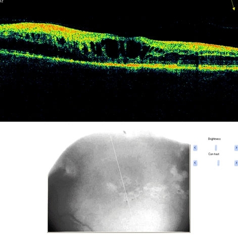

A branch retinal vein occlusion is a blockage of the portion of the circulation that drains the retina of blood. The arteries deliver blood to the retina. The red blood cells and plasma then course through the capillaries and eventually into the venous system, eventually reaching the central retinal vein. With blockage of any vein, there is back-up pressure in the capillaries, which leads to hemorrhaging and fluid leakage on the retina. Usually, the occlusion occurs at a site where an artery and vein cross. Typically, the artery crosses over the vein so that any hardening of the artery compresses the vein (which has a thinner wall). The occlusion site determines the extent or distribution of the hemorrhage, ranging from a small branch veins giving rise to occlusions involving part of the retina to a hemispheric (hemi-retinal) occlusion involving one half of the retina, to an occlusion of the central retinal vein, which involves the entire retina (when the central vein is involved, this is called a central retinal vein occlusion which is discussed below).
Branch retinal vein occlusions are by far the most common cause of retinal vascular occlusive disease. Males and females are affected equally. Most occlusions occur after age 50, although younger patients are sometimes seen with this disorder (in this age group it is often called papillophlebitis). The highest rate of occurrence is in individuals in their 60's and 70's. These disorders are similar to those for vascular occlusive disease elsewhere in the body such as stroke and coronary artery disease. Specifically, aging, high blood pressure, diabetes, and smoking are all risk factors. It is very important to find out if there is an underlying cause to any vascular occlusive disease.