
Site Design: www.hartydesign.com
Home | About Ocular Imaging | Contact | Eye Diseases | Prevention | Technology | Links | FAQs
©2010 Ocular Imaging


Ocular Imaging
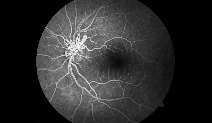
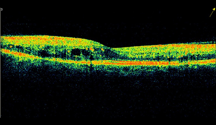
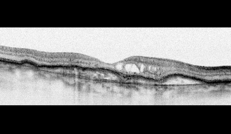
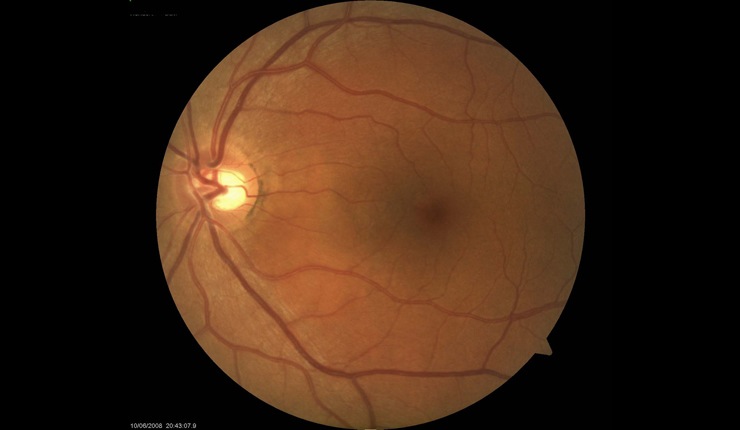
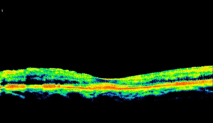
Highly reflective choroidal neovascular membrane (CNV) associated with AMD. Some residual fluid remaining at the interface of the CNV and outer retina.


Ocular Imaging
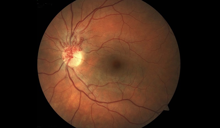
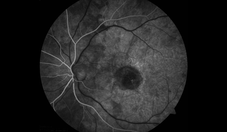
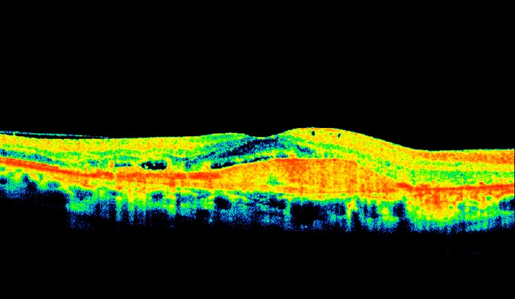
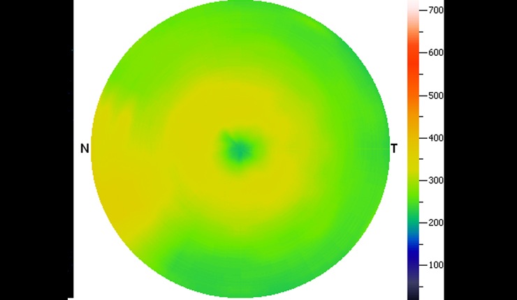
Corresponding topographic thickness map of fundus region highlighting shallow fluid temporal to the disc at the left hand side of the image (central blue/green area represents the fovea).


Ocular Imaging
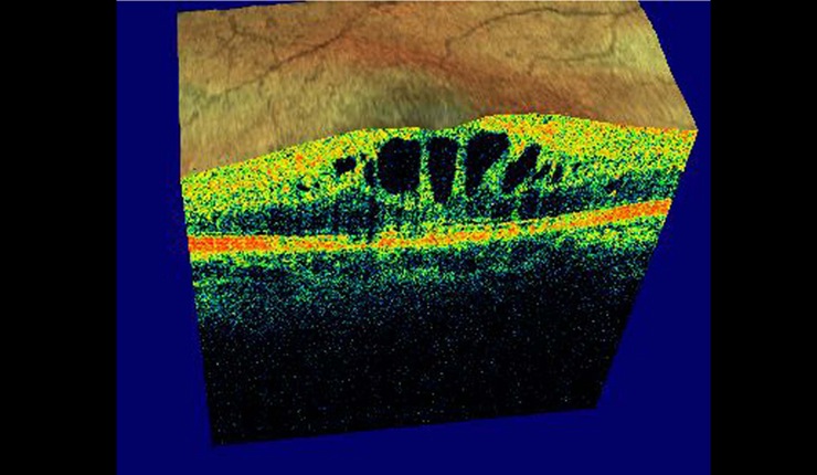
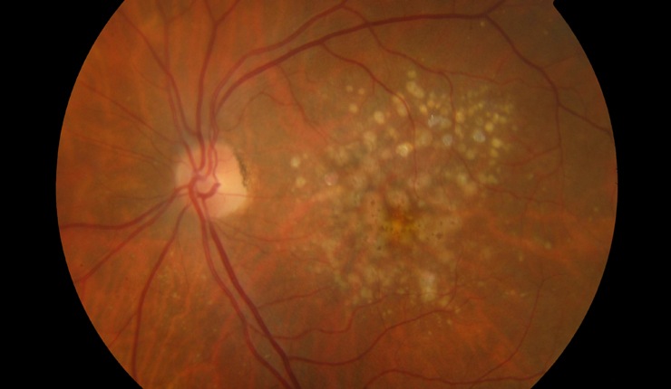
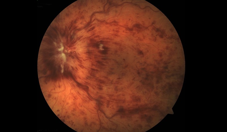
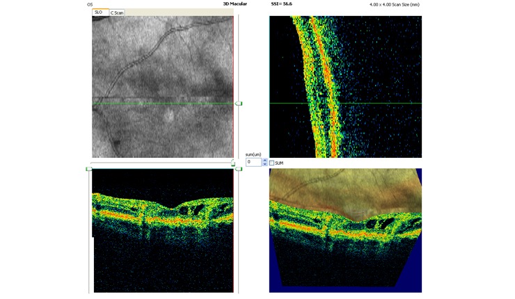
3D OCT of retinal angiomatous proliferation (RAP) showing news vessels tracking through the retina and associated intra-retinal oedema.

Ocular Imaging
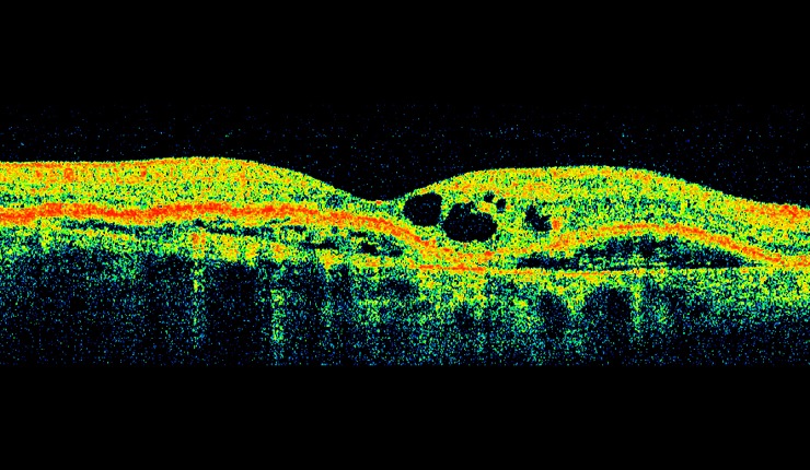
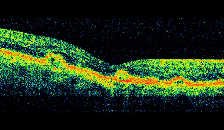
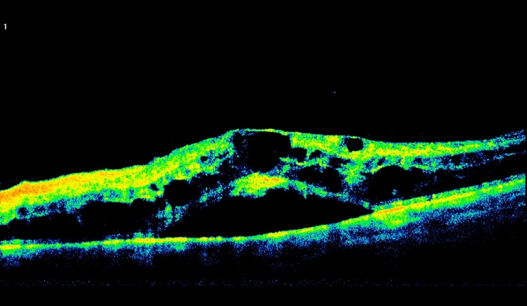
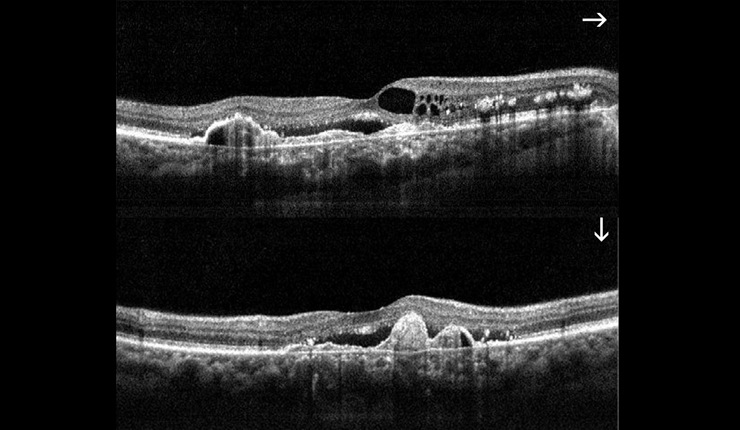
AMD. High resolution horizontal and vertical scans showing cystoids oedema, CNV, RPE proliferation and sub retinal fluid at the fovea.

Ocular Imaging
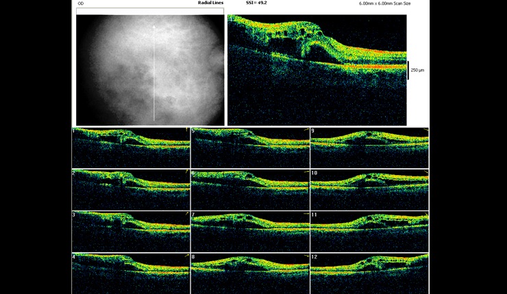
OCT radial scan display showing arcade pattern of fluid associated with branch retinal vein occlusion.

Ocular Imaging
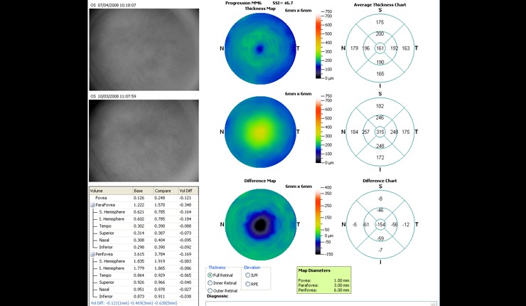
Progression retinal thickness maps showing resolution of fluid following triamcinilone treatment.